Checkov, Peanut and Molly: A Tale of Three Cats
Since November the Vets Now Referrals Glasgow team have successfully treated three very memorable feline patients. Checkov, Peanut and Molly were all diagnosed with pyothorax and referred for specialist treatment. Pyothorax is defined as pleural accumulation of purulent exudate secondary to bacterial infection. Clinical signs include increased respiratory effort, lethargy, pyrexia and inappetence. It can arise due to penetrating wounds, migrating foreign bodies, as an extension of broncho-pulmonary or other infectious diseases.
Treatment for pyothorax can be challenging yet rewarding. Checkov (5.5y MN Somali) and Peanut (7m MN Bengal) had bilateral small-bore chest drains placed whereas Molly (5.5y FN DSH) had only a single drain placed. Traditionally large-bore trocar-type drains, using pressure to insert into the pleural space or non-styletted drains placed using a “mini-thoracotomy” technique were used. Placement of large-bore thoracic drains in human medicine has been shown to have a high complication rate, in approximately 5-35% of cases. In veterinary medicine, this complication rate is reported to be even higher, in up to 58% of cases. Reported complications include pneumothorax, lung trauma, arrhythmias, haemorrhage from lacerated intercostal vessels, misplacement, failure to drain and leakage around the insertion site. Placement of small-bore chest drains is now getting more and more popular in human pleural space disease to minimize the above risks and is also considered to be more comfortable for the patient.
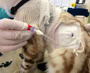
Under light sedation we placed 14g small-bore wire-guided chest drains (Chest Drain; MILA International Inc.) using a Seldinger technique. This technique is similar to that of placing a central venous line into the jugular vein and is easier and safer to perform in comparison to more invasive techniques. General anaesthesia or heavy sedation is rarely required which makes it an especially suited option for dyspnoeic patients.
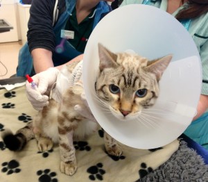
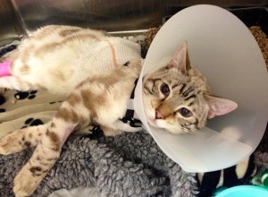
Despite the smaller gauge of the drain, we did not encounter any significant difficulties with regards to drainage of the thoracic cavity. Even the most flocculent purulent exudate did not result in chest drain failure due to obstruction. Warmed sterile saline was flushed through the drains and aspirated on a daily basis or more frequently depending on disease severity. Checkov, Peanut and Molly tolerated their thoracic drains very well and they stayed in situ for 4-5 days without any apparent discomfort. All three were hospitalised in the main ward with the necessary precautions i.e. buster collar and surgifix vest, ensuring the patients were unable to interfere with their drains.
Samples of pleural fluid were sent for cytology, culture and sensitivity. Empirical broad spectrum, combination antibacterial therapy was initiated e.g. metronidazole and cefuroxime IV initially and modified as necessary based on culture results. The most common infectious agents involved in pyothorax include aerobes and anaerobes, Pateurella spp, Actinomyces spp, Nocardia spp, Bacteroides spp, Fusobacterium spp and Clostridium spp. Long-term antibiotic therapy was continued after hospitalization for up to 6-8 weeks.
Prognosis for cats with pyothorax is fair to good with early diagnosis and aggressive medical management. Thankfully Checkov, Peanut and Molly are all currently doing well and happy at home after their treatment and hospitalisation at Vets Now Referrals.


