There had been minimal improvement after treatment with NSAIDs and physical and neurological examinations were unremarkable.
There was no obvious pain upon manipulation of the jaw, although it was difficult to assess due to the anxious behaviour of the dog during the examination.
The most likely differential diagnoses were: bilateral temporo-mandibular joint disease (TMJ); an inflammatory disorder affecting middle ear or oral cavity; masticatory muscle myositis (MMM) with an unusual presentation; and polymyositis.
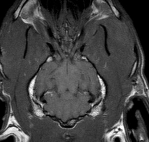
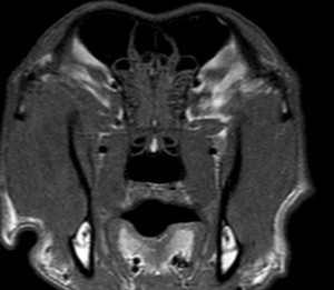
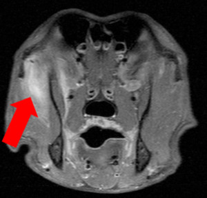
Pre-anaesthetic blood work and radiographs of the oral cavity and TMJ did not reveal any abnormalities. An MRI of the skull was then performed which revealed multiple contrast enhancing lesions in the temporal muscles.
The most severe lesion was within the left temporal muscle measuring 4cm from dorsal to ventral, by 2 cm from lateral to medial, by 3cm from rostra-caudal (see Figs. 1 and 2). These changes were consistent with a focal myositis affecting the temporal muscles (MMM or immunomediated/ infectious polymyositis)


